Wide-Angle Brain Mapping: How ULTRA Two-photon Microscopy Peers Deep
Source PublicationLight: Science & Applications
Primary AuthorsYang, Zhou, Lang et al.
Seeing Through the Keyhole
Imagine trying to watch a sold-out stadium concert through a drinking straw. You might see the lead singer’s left eye, but you miss the drummer, the bassist, and the crowd’s reaction. To understand the show, you need to see everyone at once.
Neuroscientists face this exact problem. Standard imaging tools typically trade scale for detail. They either zoom in on a few cells or zoom out to see a blurry overview of the whole organ.
Scaling Up Two-photon Microscopy
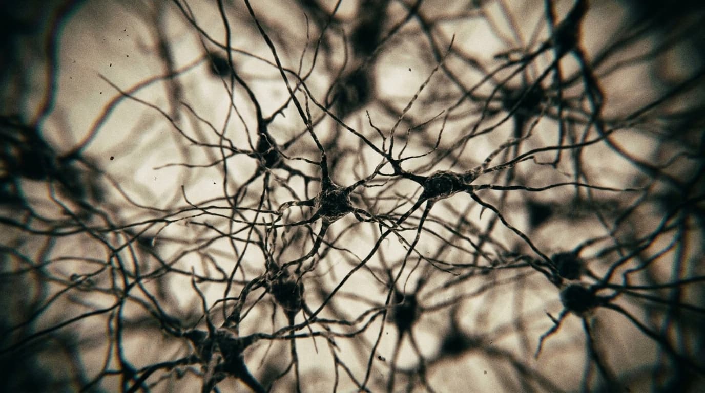
Researchers have built a system called ULTRA to break this technical bottleneck. It uses two-photon microscopy to scan an area exceeding 50 square millimetres. This is massive for a device that maintains single-cell resolution.
The team measured neuronal structures and firing patterns across a volume of 45.24 cubic millimetres. The system penetrates 900 micrometres into the mouse brain, capturing data from both superficial and deep layers.
Mapping Neural Circuits
This capability suggests that scientists can now track how distant brain regions coordinate in real-time. By removing spatial limits, the platform provides a tool to observe how complex circuits function across the entire cortex. Potential applications include:
- Tracking sensory inputs across different lobes.
- Observing deep cortical layer interactions.
- Mapping large-scale neural networks at cellular resolution.
Future work could use this to study how sensory inputs from one area influence motor outputs in another. It brings researchers closer to a complete map of brain communication.