Why Multimodal Single-Neuron Analysis is the Ultimate Upgrade for Brain Mapping
Source PublicationCell
Primary AuthorsZhao, Shi, Liu et al.
Imagine trying to understand your best friend by only looking at their Spotify playlist, or only their seat in the cafeteria, or only how they react when a surprise pop quiz is announced. You would miss the big picture. To truly understand them, you must profile their tastes, their location, and their behaviour simultaneously.
These results were observed under controlled laboratory conditions, so real-world performance may differ.
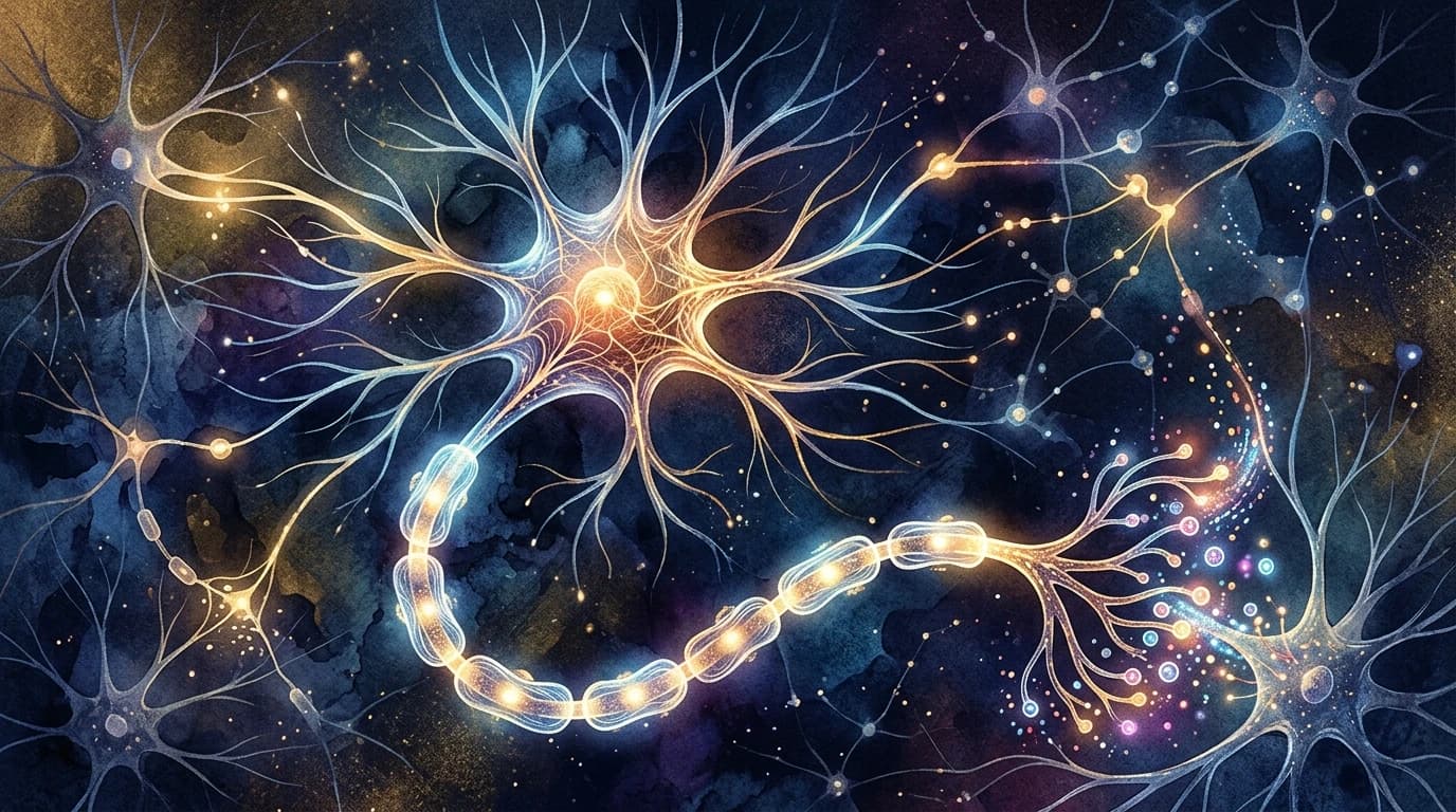
Neuroscientists have faced a similar limitation with brain cells. They could track a neuron's calcium activity, map its physical shape, or profile its active genes using imaging—but rarely all three in the exact same cell. This siloed data made it difficult to organise a clear map of how individual cells process sensory information.
A research team recently developed a platform that measures all three properties in the same neuron. They tested this on 141 visual cortex neurons in mice, tracking how calcium levels changed in response to visual stimuli, mapping their physical branches, and profiling their RNA using imaging-based in situ transcriptomics. The data revealed that physical shape and gene expression are complementary; combining them predicts a neuron's actual function far better than looking at either feature alone.
Predicting Brain Function with Multimodal Single-Neuron Analysis
This approach suggests we can no longer categorise brain cells by just one characteristic. By integrating physical structure with genetic activity, scientists can build more accurate models of the brain. While this platform is currently limited to mouse models in a lab setting, these foundational rules of the brain could eventually help researchers build incredibly detailed atlases of neural circuitry, showing exactly how complex networks are built and maintained.