The Curious Lifecycle of Neurofibrillary Tangles: A Proteomic Detective Story
Source PublicationJournal of Neuropathology & Experimental Neurology
Primary AuthorsJimenez Jaramillo, Nedderman, Ahat et al.
Is there a perverse elegance to the way our cellular machinery falls apart? We often imagine disease as pure chaos—a messy slide into disorder—but biology rarely works without a strict, albeit sometimes disastrous, logic. Even in the wreckage of a dying neuron, there is a timeline.
A recent study applies a fascinating combination of artificial intelligence and laser capture microdissection (LCM) to map this timeline. Rather than analysing bulk brain tissue, which muddles the signal, the researchers trained an algorithm to hunt specific quarry within the entorhinal cortex and hippocampus. They sought to categorise the evolution of Tau protein aggregates, separating them into three distinct phases: the early 'pretangle', the fully formed 'mature' tangle, and the spectral 'ghost' tangle.
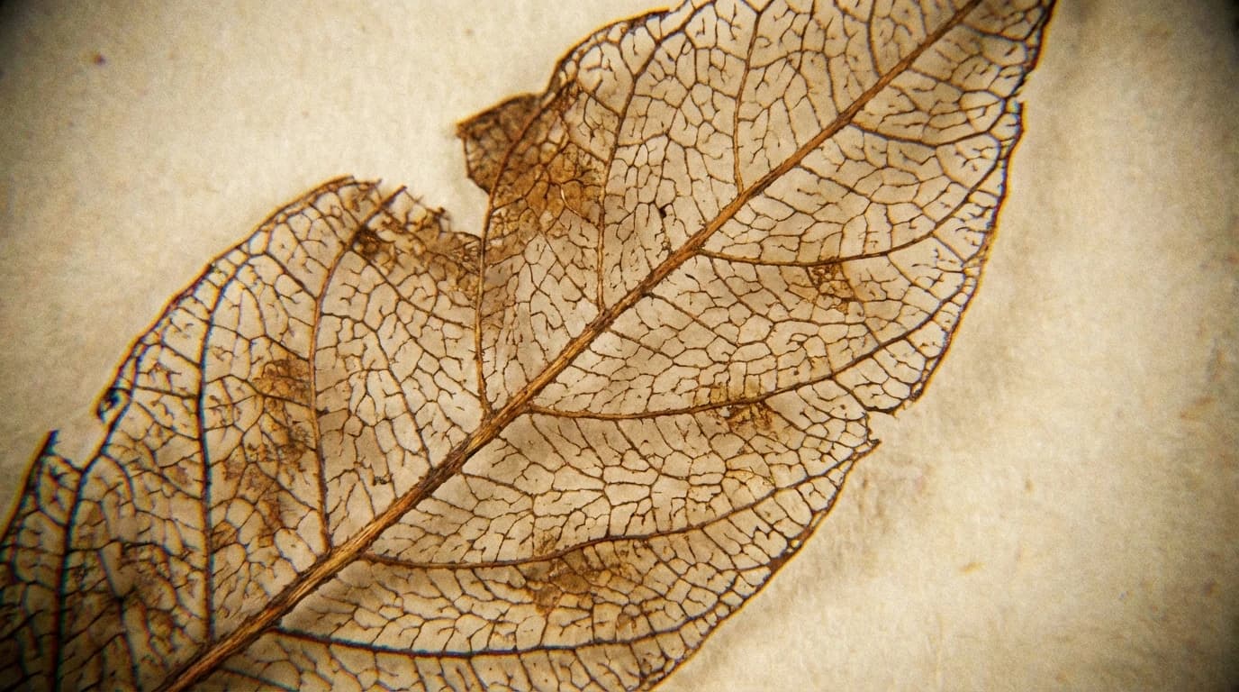
Tracing the lifespan of neurofibrillary tangles
The precision here is what matters. By physically cutting out these microscopic structures with a laser, the team could perform targeted proteomics on roughly 1,250 individual collections. What they measured was a distinct chemical arc. As the pathology progresses from the pretangle stage to the mature stage, the abundance of specific Tau signatures (notably pTau217) surges—increasing by roughly 11-fold.
Then, something curious happens. The signal collapses.
When the researchers analysed the 'ghost' tangles—the remnants left behind after a neuron has died—the levels of pTau217 dropped by up to 116-fold. The study measures this sharp reduction, but it leaves us to ponder the mechanism. Why does the protein signature vanish so completely once the cell is gone?
This invites a brief philosophical detour regarding genomic organisation. Nature rarely wastes energy maintaining structures that serve no purpose, even pathological ones. The 'ghost' tangle represents an extracellular tombstone. Once the neuron's membrane is compromised and the cell perishes, the internal environment that supported the hyperphosphorylation of Tau is lost. The sudden drop in pTau suggests that these proteins are either degraded by extracellular enzymes or dispersed into the surrounding fluid.
The data implies that our current biomarkers are catching the disease at a very specific, perhaps transient, moment of high toxicity. By distinguishing between the active 'mature' phase and the remnant 'ghost' phase, we gain a clearer view of the enemy. We are not just looking at a static scar; we are watching the wound heal, or perhaps, fester, in real-time.