How ultrasound deep learning creates sharp scans from raw echoes
Researchers reviewing medical imaging find that neural networks can process raw sound echoes into high-resolution scans at ultrafast speeds, bypassing slow mathematical calculations.
Reading level
The full story with proper science words explained.
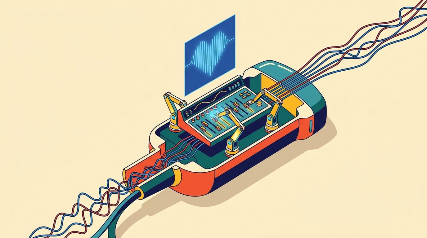
Listening to the body's echoes
An ultrasound probe listens to the human body the way our ears pick out a voice in a noisy room.
The probe fires sound pulses into tissue and records the echoes that bounce back. To turn those messy sound waves into a clear medical image, scanners rely on beamforming, a signal-processing technique that lines up and combines raw echoes from dozens of tiny sensors on the probe face.
For decades, the standard tool has been delay-and-sum. It delays the signals from different sensors so they arrive at the same moment, then simply adds them together. It is fast enough for live video, but the results can look fuzzy. Unwanted acoustic echoes, known as sidelobes, leak into the recording and clutter the screen.
The maths bottleneck
Engineers know how to sharpen these images. Advanced methods like minimum variance adjust the volume of each sensor dynamically to bring tiny details into focus.
Another method, delay-multiply-and-sum, multiplies pairs of sound waves before adding them, which suppresses background clutter. The catch is computational cost. These advanced methods require heavy matrix operations that slow scanners down to a crawl, making them impractical for standard clinical use.
Doctors must constantly choose between images that are fast or images that are clear.
Teaching networks to listen
A comprehensive review of ultrasound deep learning shows that trained neural networks can solve this trade-off.
Instead of running heavy mathematical matrix inversions, researchers feed raw radiofrequency channel data directly into neural networks. These models learn how sound waves interact across the sensor array, filtering out noise in an instant.
The results match the crisp resolution of complex algorithms while running at ultrafast speeds. One hybrid model, called ABF-MV, matched the image contrast of minimum variance beamforming while achieving frame rates of roughly 1,000 frames per second. At that speed, a scanner captures one thousand full pictures every second.
Remarkably, networks can also work with less input. In international benchmark tests, deep learning models reconstructed crisp images from a single plane-wave transmission, matching the quality of older techniques that required multiple pulses fired from different angles. Even simple 1D neural networks achieved results comparable to deeper 2D and 3D models.
Why faster processing matters
Shrinking the processing burden means clinical scans no longer require bulky, expensive computers.
By replacing heavy calculations with lightweight neural networks, manufacturers can design high-performance portable probes. Devices using fewer internal sensors could soon deliver high-contrast images directly at a patient's bedside without losing diagnostic clarity.
What we still do not know
Important hurdles remain before these algorithms reach every clinic.
Many neural networks are trained on simulated wave datasets or artificial test objects called phantoms. Scientists still do not know how reliably these models will perform across diverse human bodies, where unusual tissue textures or rare diseases might confuse the system.
Engineers must also work out how to squeeze these deep networks onto tiny chips inside ultra-portable probes without draining batteries or generating excessive heat.
Science words
- Beamforming
- The signal-processing technique used to steer, focus, and combine ultrasound wave signals to create a coherent medical image.
- Delay-and-sum
- The standard, basic beamforming method that aligns received echo waves with time delays and sums them together.
- Sidelobes
- Unwanted acoustic beams pointing off to the side of the main beam that cause image distortions and visual clutter.
- Minimum variance
- An advanced beamforming algorithm that dynamically calculates the best weights for incoming sound signals to maximize resolution.
- Radiofrequency channel data
- The raw, unprocessed electrical signals captured straight from each individual sensor element on an ultrasound probe.
- Plane-wave imaging
- An ultrafast ultrasound technique that transmits a flat, unfocused wave across the entire field of view at once.
Check it yourself
This story is based on a real research paper in International Journal of Biomedical Imaging by Hadri, Fail, Sadik. We write with AI help and check it against the paper, but the original is the final word.