Beyond the Biopsy: The Trajectory of AI in Prostate Cancer
Source PublicationScientific Publication
Primary AuthorsNair RP, Du W, Mei L, Koga S.
For decades, diagnosing prostate tumours has relied on manual, subjective reviews of biopsy slides, leading to varied interpretations and unnecessary procedures. Now, the integration of AI in prostate cancer research offers a clear path past this diagnostic bottleneck.
These results were observed under controlled laboratory conditions, so real-world performance may differ.
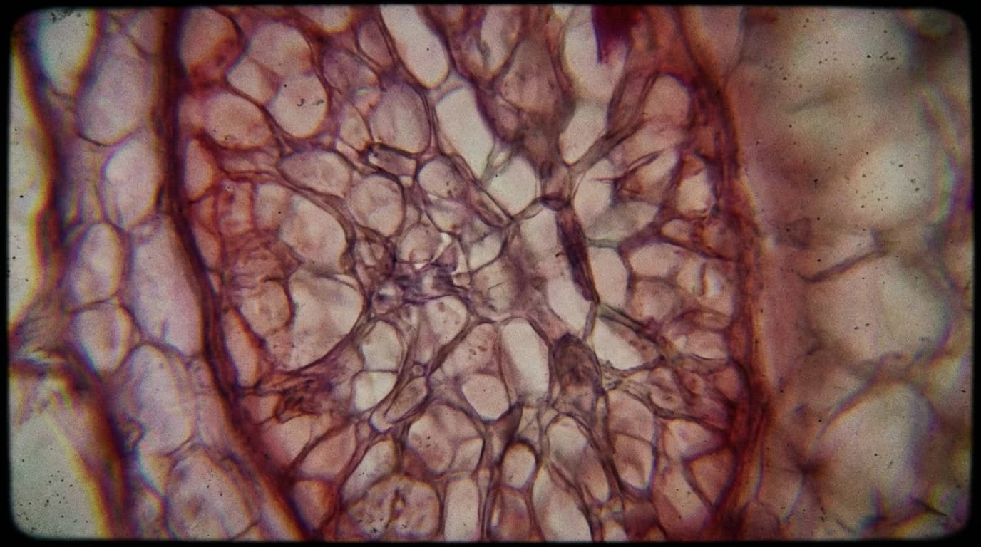
Assessing tumour severity traditionally depends on human eyes examining tissue samples under a microscope. This manual process takes considerable time and leaves room for human error.
Machine learning models have recently demonstrated expert-level potential in retrospective studies to address these inefficiencies. While the field still needs to move toward clinical use, these algorithms are learning to spot microscopic anomalies with the accuracy of seasoned specialists, potentially reducing the need for invasive tests.
Current Capabilities of AI in Prostate Cancer
Recent studies measured the performance of deep learning algorithms against human pathologists. The data shows these tools can assign Gleason grades and identify malignant cells with high accuracy.
Furthermore, the models successfully measured prognostic markers, such as Ki-67, and flagged complex cases for human review. The technology even suggests molecular alterations and lymph node spread directly from standard histology images.
This capability could provide a highly cost-effective alternative to traditional laboratory assays. Natural language processing and large language models are further expanding this potential.
These tools extract complex information from clinical notes and translate it to enhance patient education, expanding the technology's utility far beyond just image analysis.
The Next Decade of Diagnostics
Looking ahead to the next five to ten years, this technology could drastically alter patient care. Multimodal models will likely combine digital pathology with a patient’s specific clinical data to recommend personalised treatments.
The implications for the next decade are substantial. Rather than relying solely on visual tissue inspection, oncologists could use these systems for improved outcome prediction and risk assessment before a scalpel ever touches the patient.
However, the transition to widespread clinical use requires overcoming current hurdles. Most models currently suffer performance drops when tested at new medical centres.
These drops typically occur due to differences in patient populations and slide preparation techniques. To integrate these tools safely, the medical community needs:
- Prospective, multicentre validation trials to ensure reliability.
- Strict standardisation for preanalytical and analytical processes.
- Clear reporting guidelines for human oversight and model failures.
Emerging systems, such as transformer-based image models and vision-language tools, could improve how well these algorithms generalise across different hospitals. Self-supervised pretraining may also help these systems learn from smaller datasets.
By addressing these technical variations, artificial intelligence may soon offer a highly precise standard of care. This shift suggests a future where fewer patients undergo unnecessary biopsies, and those who require treatment receive highly targeted, data-driven therapies.